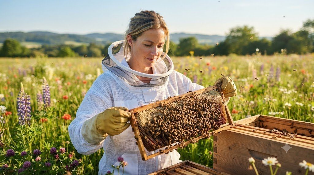
Every time an apiarist lifts a brood frame out of the hive and scans the comb surface, they are acting as the frontline veterinary epidemiologist for their colony. A healthy brood nest displays a dense, contiguous mosaic of pearly-white, plump C-shaped larvae glistening in pools of royal jelly, surrounded by convex, uniform, golden-tan cappings with minimal empty cells (‘shotgun brood pattern’). When bacterial, fungal, viral, or microsporidian pathogens invade the brood chamber, that visual symmetry breaks down quickly.
Distinguishing accurately between American Foulbrood (Paenibacillus larvae)—a lethal, spore-forming regulatory plague that requires immediate quarantine and destruction of contaminated comb—and European Foulbrood (Melissococcus plutonius), Chalkbrood, Sacbrood, and Nosema ceranae is the single most critical diagnostic responsibility in beekeeping.
American Foulbrood (Paenibacillus larvae): Endospores and the Ropiness Test
American Foulbrood (AFB) is caused by the Gram-positive, rod-shaped, endospore-forming bacterium Paenibacillus larvae. Only young worker, queen, or drone larvae less than 53 hours old are susceptible to infection (with Newly hatched <24-hour larvae requiring fewer than 10 spores ingested in contaminated brood food to contract a fatal infection!). Once inside the larval midgut, the spores germinate into vegetative rods that proliferate rapidly, penetrating the gut epithelium into the hemocoel and killing the larva AFTER the cell has been capped with wax (during the prepupal or early pupal stage).
Why is AFB so terrifyingly persistent? Because as the liquefied prepupa dries down into a brittle, black, tightly glued wafer along the lower wall of the cell (an AFB scale), the bacteria sporulate—producing roughly 2.5 billion endospores per single dead larva! These endospores possess a multi-layered protein coat that remains infectious for 40 to 70+ years, surviving boiling water at 100°C, freezing, alcohol, and household bleach. When innocent house bees attempt to clean out the dead scale, they contaminate their mouthparts and honey stores; worse, when the colony weakens and dies, neighboring hives rob out the contaminated honey and spread AFB across the entire 3-kilometer radius!
Executing the Field Matchstick Ropiness Test and Holst Milk Test
When inspecting a patchy ‘shotgun’ brood frame, look closely for sunken, concave, dark, greasy-looking cappings perforated with irregular pinholes. To perform the classic field Ropiness Test:
- Insert a rough wooden toothpick, dry grass stem, or matchstick through a sunken perforated capping into the caramel-brown decaying prepupal mass at the bottom of the cell.
- Stir the contents gently and withdraw the stick slowly away from the cell face.
- If the decayed brown mass stretches out in a uniform, elastic, gluey thread 2.5 to 5.0 cm (1 to 2 inches) long before snapping back like warm melted mozzarella cheese, and emits a sulfurous, decaying animal-glue odor, suspect American Foulbrood immediately! Looking down at the lower cell wall at a 45-degree angle with sunlight over your shoulder may also reveal a dried pupal tongue protruding upward from the dark scale toward the roof of the cell—a pathognomonic sign unique to AFB.
- Immediately burn or bury the test toothpick, wash your hive tool and hands with rubbing alcohol followed by mechanical scrubbing with soap and water (alcohol kills vegetative bacteria, while mechanical soap scrubbing washes off spores; never use your contaminated smoker or gloves on another hive!), and contact your State Apiary Inspector or run a Vita AFB Lateral Flow Immunochromatographic Diagnostic Strip.
American Foulbrood is a legally reportable disease across most US states, Canadian provinces, UK, EU, and Australasia. While antibiotics like Oxytetracycline (Terramycin) or Tylosin (under a licensed veterinarian’s VFD prescription) suppress vegetative rods and mask visible symptoms, antibiotics DO NOT KILL AFB SPORES!The moment antibiotic feeding stops, dormant spores in the comb and honey germinate and trigger a relapse—which is why burning infected frames/comb (or gamma irradiation) remains the definitive biosecurity cure.
Differential Diagnostic Matrix of Honey Bee Brood & Adult Pathologies
Use this clinical reference table to differentiate between the five major brood and adult digestive diseases encountered during field inspections.
| Disease & Causative Agent | Stage at Death | Capping Appearance | Larval Color & Consistency | Matchstick Rope Test | Definitive Field & Lab Treatment |
|---|---|---|---|---|---|
| American Foulbrood (Paenibacillus larvae) | Capped Prepupa / Pupa | Sunken, greasy, dark, perforated holes | Coffee-brown to black brittle tight scale | POSITIVE (Ropes 2.5–5.0 cm elastic thread) | Report to Inspector; burn frames or gamma irradiate |
| European Foulbrood (Melissococcus plutonius) | Uncapped Coiled Larva (Days 4–5) | Uncapped open cells (mostly) | Yellowish-tan, twisted, visible tracheal lines | NEGATIVE (Watery/granular; <1 cm coarse) | Requeen (brood break) + 1:1 syrup feed; VFD if severe |
| Chalkbrood (Ascosphaera apis – Fungus) | Stretched Larva just after capping | Uncapped by nurses or perforated | Hard white or gray-black chalky mummies | NEGATIVE (Hard dry solid piece of chalk) | Improve ventilation/sunlight; requeen with hygienic stock |
| Sacbrood Virus (SBV – Iflavirus) | Stretched Prepupa in capped cell | Uncapped or pinhole openings | Tough fluid-filled sac; darkened Gondola head | NEGATIVE (Lifts out intact like water balloon) | Remove worst frame; requeen if >5% brood affected |
| Nosema ceranae / N. apis (Microsporidia) | Adult Bees Only (Midgut epithelium) | Brood cappings unaffected | Swollen pale/grayish adult ventriculus (gut) | N/A (Microscopic oval spores at 400x) | Ensure sunny dry winter site; young combs; Fumagillin |
European Foulbrood (Melissococcus plutonius): Nutritional Stress Recovery
Unlike AFB, European Foulbrood (EFB) is caused by a non-spore-forming Gram-positive lanceolate coccus bacterium, Melissococcus plutonius (frequently accompanied by secondary invaders such as Paenibacillus alvei and Enterococcus faecalis). EFB bacteria multiply inside the midgut of open coiled larvae, competing directly with the larva for food. Consequently, EFB classically erupts in early spring when colony brood nests expand faster than nurse bees and incoming pollen can feed them—or during stressful migratory pollination in cranberry and blueberry bogs.
Because M. plutonius starves the larva before metamorphosis, EFB kills the larva while the cell is still OPEN and uncapped (around Days 4–5). Infected larvae lose their plump pearly-white sheen, twist corkscrew-style out of position along the cell walls, turn translucent yellowish-brown revealing a prominent network of white silver tracheal breathing tubes, and dry into rubbery scales that house bees can easily pull out and discard. In mild-to-moderate cases, caging or replacing the queen to induce a 14-day brood break while feeding warm 1:1 sucrose syrup and protein patties allows hygienic nurse bees to eject every infected larva before the new queen begins laying.
Nosema apis vs. Nosema ceranae: Microscopic Spore Counting at 400x
Two obligate intracellular microsporidian fungal parasites infect the epithelial cells of the adult honey bee midgut (ventriculus): historic Nosema apis (which causes late-winter dysentery—brown fecal streaking across the front of the hive when bees are confined during long cold spells) and emergent Nosema ceranae (which causes ‘dry nosema’—year-round energetic starvation, suppressed vitellogenin, premature foraging, and sudden spring depopulation without obvious fecal streaking). Because visual field symptoms of N. ceranae are subtle, accurate diagnosis requires grinding the abdomens of 25 returning forager bees in 25 mL of distilled water (1 mL per bee), placing a droplet on a hemocytometer slide under a 400x phase-contrast compound microscope, and checking for bright, rice-shaped oval refractive spores measuring $4.5 \times 2.2$ microns. Maintaining sunny, dry overwintering yards, rotating out dark brood combs every 3 to 4 years, and ensuring rich autumn pollen nutrition keeps Nosema spore counts safely below the 1-million-spores-per-bee clinical threshold.
Hygienic Biosecurity Protocols and Comb Rotation Schedules
In medicine, prevention is always infinitely cheaper than cure; in apiculture, biosecurity is the difference between a thriving 20-year apiary and a total regulatory burn order. Because bacterial endospores (Paenibacillus larvae), fungal ascospore cysts (Ascosphaera apis), and microsporidian spores (Nosema ceranae) accumulate progressively inside dark, multi-year brood comb cocoons and stored honey, implementing a strict 5-Point Apiary Biosecurity Protocol eliminates 95% of infectious disease transmission:
- Never Purchase Used Woodenware or Dark Drawn Comb of Unknown Origin: Bargain estate-sale hives with black combs are the #1 vector for introducing dormant 40-year-old American Foulbrood spores into a clean bee yard.
- Never Feed Store-Bought Imported Honey to Bees: Commercial supermarket honey frequently contains sub-clinical loads of Paenibacillus larvae spores that are completely harmless to human digestion (except infants <12 months due to Clostridium botulinum) but fatal to honey bee larvae. Feed exclusively pure white cane/beet sucrose syrup or your own verified disease-free honey.
- 4-Year Brood Comb Renewal Cycle: Brand-new brood frames with the installation year written in indelible marker on the top bar (`2025`, `2026`). Each spring, shift the darkest, heaviest 2 or 3 brood frames to the outer edges of the box (and cycle them out once brood emerges) while inserting 2 to 3 fresh foundation frames into the edges of the brood nest, replacing 100% of brood combs every 4 years.
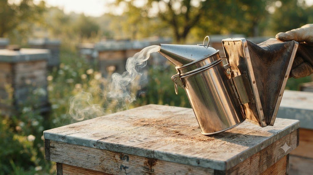
- Flame-Scorch Hive Tools Between Yards: Propolis and honey stuck to the blade of your steel J-hook hive tool can transfer AFB spores from a sick hive to 10 healthy hives in a single afternoon. Scrape your hive tool clean, plunge it into the glowing coal bed of your lit smoker for 30 seconds, and wipe with 70% isopropyl alcohol or a 10% bleach solution between yards.
Clinical Diagnostic Inspection Workflow and Lab Sampling Protocols
Translating honey bee pathobiology, endocrinology, and pheromone chemistry into thriving colonies requires a systematic, repeatable Clinical Brood-Nest Inspection Protocol. Rather than pulling frames randomly from the middle of the hive (which risks rolling the queen between tight combs), always start by removing the outermost honey/pollen frame (#1 or #10) to create a wide working gap, then inspect frames sequentially toward the center while auditing five biological biomarkers.
| Biological Biomarker | Healthy Colony Baseline | Subclinical Warning Sign | Acute Pathological Crisis | Immediate Corrective Intervention |
|---|---|---|---|---|
| Egg & Young Larval Ring | Single upright eggs centered on floor; rich pool of white jelly | Dry larvae with minimal jelly (Nutritional stress) | Multiple eggs plastered on cell walls (Laying workers) | Feed 1:1 syrup + polyfloral pollen patty; unite laying workers w/ queenright nuc |
| Capped Brood Symmetry | >90% contiguous convex tan caps; 5–8% heater shafts | 15–25% spotty empty cells; occasional chewed pupae | Sunken greasy perforated caps; ropy brown scales (AFB) | Run alcohol mite wash & matchstick rope test; isolate hive tool |
| Varroa Mite Load (300 Bees) | < 1% (<3 mites/300 bees) in spring & autumn | 2% to 3% (6–9 mites/300 bees) in summer | > 4% (12+ mites) + bees with shriveled wings (DWV) | Apply temperature-matched IPM (Formic Pro, Thymol, or Oxalic + brood break) |
| Adult Bee Gut & Demography | Plump fuzzy nurses; clean entrance; active pollen loads | Slow spring buildup; high forager mortality | Dysentery streaking; crawling bees w/ K-wings (Nosema/CBPV) | Check 400x wet mount for Nosema spores; requeen & rotate dark comb |
| Acoustic & Pheromonal Tone | Calm low 180–220 Hz hum; cohesive frame carpet | Elevated roaring pitch when smoked; empty cups | High-pitched queenless roar; fanning without homing | Insert test frame of young eggs/larvae from healthy hive to verify queen status |
The ‘Test Frame of Eggs’ Assay: Definitive Queenlessness Diagnosis
What should you do when you open a hive in mid-summer, see zero eggs and zero young C-shaped larvae, hear a loud roaring hum, yet cannot tell whether the colony is truly queenless OR whether a slim, unmated virgin queen is hiding somewhere on the combs? Never blindly drop a $45 mated queen cage into a hive that might harbor a hidden virgin queen—the resident virgin will immediately sting the caged queen through the mesh!
Instead, perform the foolproof Test Frame Biological Assay:
- Go to a healthy, booming queenright colony in your yard, select a frame containing hundreds of fresh upright eggs and newly hatched 24-hour-old larvae, brush 100% of the adhering bees back into their parent hive, and mark the top bar of this test frame with a thumbtack.
- Insert this frame of fresh eggs right into the center of the suspect colony’s brood nest and close the hive.
- Read the Result Exactly 72 Hours Later: If the colony was truly queenless, the desperate nurse bees will have pulled 5 to 15 unmistakable emergency queen cells directly over the young larvae on your test frame! You can now either let them finish one prime cell OR pinch the emergency cells and introduce a mated queen with 100% confidence. Conversely, if zero queen cells are pulled on the test frame after 72 hours, a virgin (or newly mated queen about to start laying) is already present inside the hive!
In the United States and Canada, state apiary programs and the USDA-ARS Bee Research Laboratory (Beltsville, Maryland) provide free clinical microscopy and molecular diagnostics for suspected brood and adult bee diseases! To submit a suspicious brood sample, cut a 4×4-inch square of affected comb containing dead larvae/scales, wrap it in a dry paper towel or porous newspaper inside a cardboard box (NEVER wrap comb in plastic bags or aluminum foil, which causes saprophytic mold growth that ruins lab analysis!), and mail with your apiary contact form.
Frequently Asked Questions: Pheromones, Mites, and Brood Pathology
Does an alcohol wash kill the 300 nurse bees—and is a powdered sugar roll just as accurate?
While an alcohol wash (or Dawn ultra-dish-soap water wash) sacrifices ~300 nurse bees out of a colony of 45,000 bees (where the queen lays 1,500 new eggs every single day—meaning those 300 bees represent just 4.8 hours of the queen’s daily egg output!), comparative field validations by Dr. Randy Oliver show that agitated alcohol/soapy-water washes recover 98% to 100% of phoretic mites consistently. In contrast, a powdered sugar roll frequently undercounts mites by 30% to 50% on humid days or when performed with coarse sugar, giving beekeepers a false sense of security that can cost the entire colony of 45,000 bees in winter.
How can I tell the difference between laying worker drone brood and a drone-laying queen?
Both produce raised, bullet-shaped drone cappings inside worker-sized cells, but their egg-laying patterns are completely different! A failing or unmated drone-laying queen has a long abdomen that reaches the bottom of the cell, so she still lays 1 single unfertilized egg centered neatly on the cell floor in a cohesive patch. In contrast, laying workers have short abdomens that cannot reach the bottom of a deep comb cell; they lay 5 to 15 eggs chaoticly plastered along the vertical side walls of each cell, often depositing eggs right on top of stored bee bread (pollen) cells!
Why do bees throw white pupae out on the landing board after a cold spring night?
If a fast-expanding colony stretches its brood nest across 7 frames during warm April days and a sudden -3°C (26°F) freeze strikes overnight, the cluster must contract tightly toward the center to keep the core at 34.5°C. The outermost ring of brood left outside the contracted cluster mantle suffers Chilled Brood. Unlike infectious bacterial foulbrood, chilled brood affects a neat concentric outer ring of all ages simultaneously, turns gray-black without ropiness or foul odor, and hygienic house bees rapidly uncap and drag the dead white/gray pupae out onto the front porch within 48 hours as weather warms.